|
|---|
 Industrial Group of the BCA
January 1997 - techniques
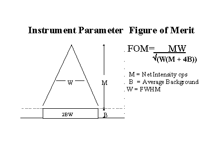
SPECIAL INTEREST GROUPSThe Industrial Group committee continues to look for ways in which our crystallography community can mutually encourage one another's expertise. One of the strengths of the Group is in our range of interests across industries and techniques which most of us find very stimulating. Sadly, the value of this cross-fertilisation is not always so apparent to our employers. The specialist seminars and workshops that are organised from time to time should have a more obvious relevance. Through these meetings, many practitioners are acquainted with the work of other laboratories and know where to turn for advice when stuck. The committee feels that the time is right to promote more cohesion within groups of practitioners who have a common specialist interest. The first such group has already been established, comprising those with an interest in pharmaceuticals. The group met last year with the BCA and is to meet again this March under the auspices of the Industrial Group. The frequency and type of meetings appropriate to such a Special Interest Group (SIG) will largely depend on the felt needs of the members, but it is expected that round-table discussions would be more common than formal presentations. SIG's could meet on their own or associated with a regular Spring or Autumn BCA meeting. To take this idea forward, we now need to know the specific interest areas which would be you would find most valuable. Some are fairly obvious, e.g. we hope to establish a non-ambient group on the basis of the workshop planned for June. I took the opportunity at the 1996 Harwell Autumn Meeting to conduct a straw poll of interests. With about 35 present, the audience was asked for a show of hands in response to a list of technique and materials interest areas. Deft counting by our Chairman produced these figures: Survey ResultsNo. Techniques 20 Texture 11 Residual Stress 10 Non-ambient 14 Line Broadening 14 Rietveld 4 Small Angle Scattering 8 Reflectometry / Grazing Incidence 1 Topography 7 Amorphous Scatter 6 Structure Determination 14 Lattice Parameters & Indexing No. Materials 12 Structural Materials(metals, ceramics, etc) 2 Electronics 5 Chemicals 7 Polymers 10 Minerals Most of those who were there felt that they would like to be involved in at least one such group. What do you think?Do you feel strongly about the need for a particular group? Have you suggestions for topics to start any such meetings? Do you think the whole idea is misguided? Please send me your views. E-mail is probably simplest [email protected] ([email protected]) or see my address and phone number in the committee list.Steve Norval Round Robin UpdateThe Instrument Sensitivity Round Robin exercise is almost complete. Thirty data sets have been evaluated and the four outstanding are promised by the end of January. A report to all participants will be issued soon after. The conclusion of the exercise will be a workshop at the Leeds spring meeting with Ron Jenkins as the main speaker. A copy of Ron's new book "Introduction to X -ray Powder Diffractometry" is included in the fee which considering that the UK price is £65 makes the workshop extremely good value for money. Overall the UK data is worse than the USA data so there is some room for improvement. From the US data Ron derived a Figure of Merit (FOM) to test instrument sensitivity - it is fully explained in his new book. I have taken the data from Test 1c of the UK data to give participants a benchmark to test their instrument sensitivity against the rest. The data from 10 and 46 are from the same instrument with the 46 data set obtained with a new X-ray tube of the same specification.
If you want to use this FOM as a guide for testing your instrument parameters the following method should be followed. The data is measured using your usual slits, 45 kV and max mA. The NBS SRM1976 Alumina plate is scanned from 50 to 55 degrees (Cu radiation) using 0.02 degree st eps and 4 second per step. The intensity of the 52.5 peak is measured in counts per second (cps) and Full Width Half Maximum (FWHM) in degrees of 2 theta of the alpha1 peak. An average of the background from 51.0 to 51.18 and 54.0 to 54.18 in cps is used. All the data are extracted manually from the raw data file. The larger the FOM the better the performance. The NBS 1976 plate can be obtained in the UK from Bureau Analysed Samples Tel 01642 300500. The FOM test is also useful when adjusti ng the parameters of an individual instrument to maximise its sensitivity. Calculating the FOM
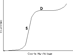
Some early conclusions can be made.: There is a wide spread of performance, many instruments have angular displacement problems which are easily overcome, countrates are lower than US and intensities more variable. Perhaps routine checks should be more widely used to ensure that equipment performs to manufacturers specification and that X-ray tube degradation is monitored. Routine checks should monitor overall or angular dependent loss in intensity or shifts in 2 theta values and changes in peak shape. Perhaps you should ask yourself a few questions. How old is my X-ray tube? When did I last check my intensities? Dare I run the FOM test on my instrument? Why haven't I got my own SRM1976? Can I afford to miss the workshop? For further details, contact Dave Taylor [email protected]Dave Taylor Help! Proportional CountersThe first of an occasional series of articles raising points which may be of interest, and which will perhaps generate discussion of how and why we should set up procedures to ensure the X-ray instrument is performing correctly. This article from Trevor raises an inte resting set of questions and a possible answer is given below. How do you set up your counters?Having recently had difficulties with a proportional counter on a diffractometer, I have started thinking about how I set it up and why. This has raised a number of questions about custom and practice. Conversations with other Industrial Group members has brought responses such as - 'I leave it to the service engineer' or 'I have always done it such-and-such a way'. I am in thi s article purposefully avoiding introducing scintillation counters into the controversy. Setting up the counter involves selecting suitable values for high voltage (HT), amplifier gain and lower and upper pulse height analyser (PHA) levels. As each affects the others, it is important that they are done in the correct sequence. This article considers only the first step of setting the counter HT. This involves progressively increasing the HT setting with a constant stream of X-rays incident on the counter. During this procedure, it is particularly important to set the PHA window as wide as possible (commensurate with removing background noise) otherwise false detector curves will be obtained.
The figure shows the output count rate from a proportional counter as the HT applied to the counter anode is increased from zero to 2000 volts or so. There appear to be two main traditions for selecting the op timum HT setting which I will call the 'diffraction' and the 'spectrometry' methods. I was brought up in the diffraction tradition in which the beginning of the plateau region (marked D) is selected, on the grounds that output signal is then at a maximum and the output signal will be insensitive to drift in the HT supply. Colleagues operating X-ray spectrometers choose a point in the middle of the linear slope (marked S) wi th the justification that this gives true proportionality between input X-ray energy (or wavelength) and output pulse amplitude (hence the term 'proportional counter'). One might conclude that the needs of diffraction and spectrometry are different, thus justifying different settings. However, one diffractometer manufacturer has issued a handbook indicating selection of point S as the correct operating position. Another recommends setting the PHA window first and then adjusting the HT for maximum counts. It is not clear what point on the graph this selects. Hopefully other readers will have views on this issue and I am sure that the editor will be happy to offer them space. Some literature references to confuse you further:- 1. U W Arndt - Chapter 7 of X-ray Diffraction by
Polycrystalline Materials (Peiser, Rooksby & Wilson, IOP
1955) 2. V E Buhrke - Chapter 3 of Handbook of X-rays (E F Kaelble)
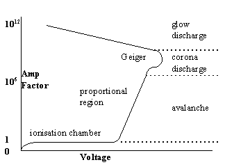
McGraw Hill 1967. 3. R Jenkins & JL DeVries - Chapter 3 of Practical X-ray Spectrometry, Macmillan 1970. Details the various regions of the HT graph, describing the S region as 'Proportional', the knee of the curve as 'limited proportionality' and the plateau as the 'Geiger region'. Trevor Carter Editor's Reply:Cullity (1) provides the best description, in particular the section on proportional counters clarifies the queries raised by Trevor. The graph shown above is reproduced in many publications, but it relates the applied voltage to observed pulse height. A more instructive curve is amplification factor vs voltage, as shown in Cullity:
From this, the amplification factor remains at 1 (ionisation chamber region) until a critical voltage is reached where avalanche occurs, and this leads to the proportional region, where the pulse height is proportional to the energy of the incident radiation. Beyond this, corona ( Geiger region ) and then glow discharge occurs, and proportionality is lost. For diffrac tion work, therefore, the position shown by Trevor (D) is correct, Cullity suggests a point 100volts along the plateau, where the counter is working in a proportional mode (i.e. below the Geiger region ) but where HT variation will not affect the pulse height. This latter consideration is less of a problem in these days of solid state electronics. The corona discharge region lies at the far end of the plateau, and results in the sharp rise of response with voltage. The issues raised by Trev or are of interest - how many of us actually check the conditions under which we are operating? Certainly my understanding is that the PHA window should be 0-100%, with the goniometer set to a peak giving 2,000 to 4,000cps, to find and set the operating voltage on the plateau. The plateau voltage will vary with the tube target used. Then a narrow (2%) window is selected to give a pulse height distribution curve, from which the upper and lower window levels may be chosen. The problem these days with "black box" electronics is that it is not always clear what conditions have actually been chosen by the hardware if an automatic set up procedure has been used. If there are any other thoughts or comments on this subject, please let me know, especially in these days of standard methods and procedures. (1) Cullity, B. D. Elements of X-ray Diffraction 2nd Ed. Addison-Wesley 1977 pp 199 - 216 Bruce Fox Spurious peaks from SellotapeI struggled for a some time with a problem peak which was almost in the correct position for a beta phase of a particular polymer, but correlation of the X-ray data with polymer processing conditions proved impossible. By chance I discovered that, if I inadvertently stretched the double sided Sellotape holding the sample (we were not interested at that stage in the amorphous content, so the tape covered the whole of the underside of the thin sample), th e "beta" phase peak appeared. A little investigation showed that some makes of sticky tape give a crystalline phase when only lightly stretched. So beware! Any similar observations or tips would be gratefully received. Bruce Fox |

|